Female Pelvis Anatomy Muscles / The Muscles That Control the Pelvic Floor | PeriCoach - Overview of the anatomy, location and function of the pelvis and perineum.
Female Pelvis Anatomy Muscles / The Muscles That Control the Pelvic Floor | PeriCoach - Overview of the anatomy, location and function of the pelvis and perineum.. Pelvic skeleton includes two hip bones, sacrum and coccyx. It's about 2 finger widths wider and 2 finger widths shorter than a male pelvis. In terms of comparative anatomy the human scapula represents two bones that have become fused together; The hip joints (acetabulofemoral joint) are joints located between the head of. Vides a discussion of the contemporary understanding.
Boundaries of the pelvic outlet (anterior, lateral and posteri… male or female: The female pelvis, which accommodates the birth canal, is larger and wider than the male. The best way to memorize and retain information is by spaced repetition. The pelvic diaphragm comprises of the two paired muscles and their fasciae; Included within the chart are gorgeous illustrations of the pelvic diaphragm, sphincter muscles, gluteus maximus.
Post Pregnancy Bodies - Pelvic Floor - Healthy Post Natal Body from www.healthypostnatalbody.com Female pelvis anatomy | free axial cross sectional anatomy of female pelvis. The hip joints (acetabulofemoral joint) are joints located between the head of. Boundaries of the pelvic outlet (anterior, lateral and posteri… male or female: The muscles of the levator ani are important supportive muscles for the midline organs of the pelvis. The muscles of the pelvis, hip and buttock anatomical chart shows how each muscle in this area of the body works with the others, and the various minor systems within the major ones. 4 write in a tabulated form origin, insertion levator prostate(in males) /pubovaginalis (in females): The pelvic diaphragm comprises of the two paired muscles and their fasciae; Above the pelvic brim and has no obstetric importance.
Included within the chart are gorgeous illustrations of the pelvic diaphragm, sphincter muscles, gluteus maximus.
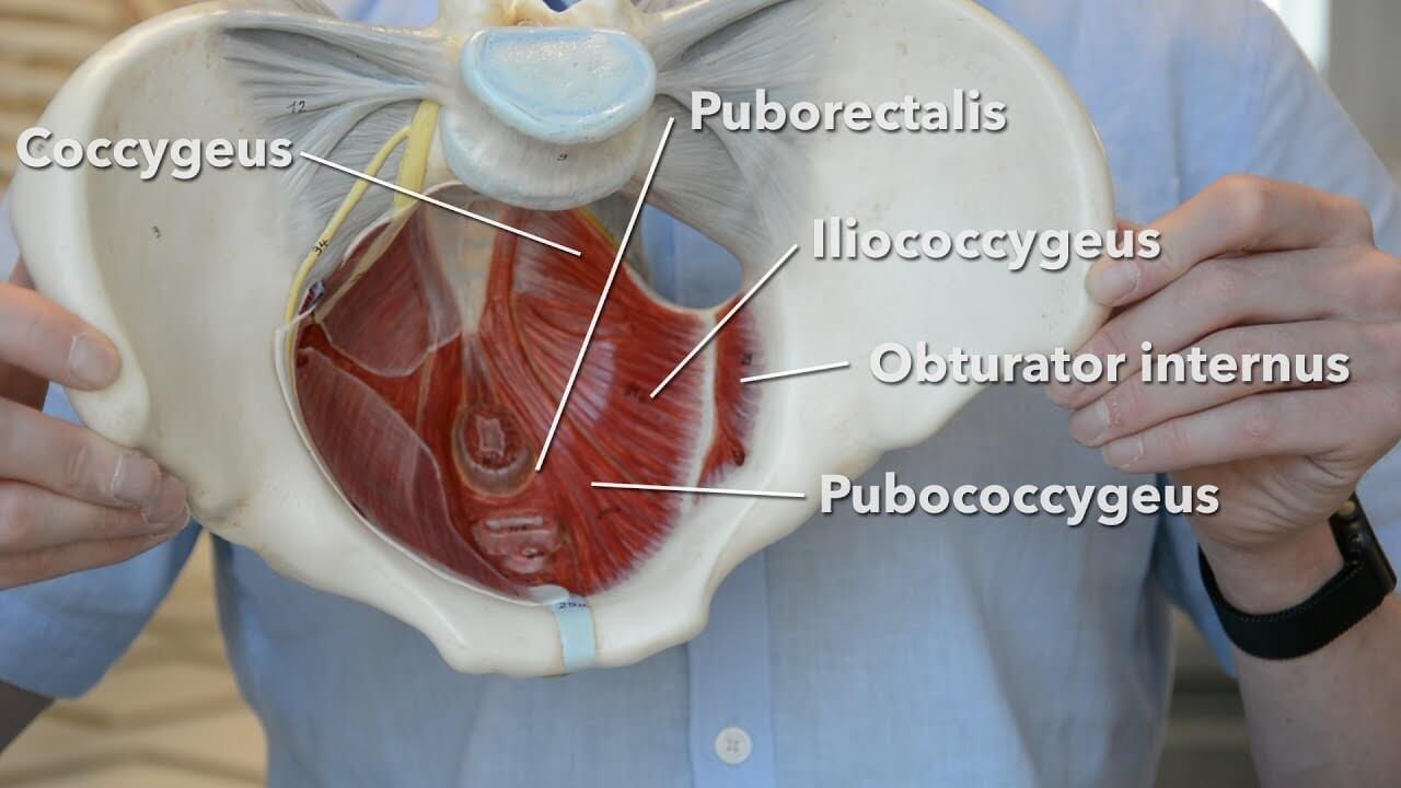
4 write in a tabulated form origin, insertion levator prostate(in males) /pubovaginalis (in females): Within the cervix itself, the anatomy is subdivided into the endocervix and the exocervix or ectocervix. There are many muscles that form the pelvic floor, including puborectalis, pubococcygeus, iliococcygeus. These muscles, including the gluteus maximus and the hamstrings, extend the thigh at the hip in support of the body's weight and propulsion. We'll go over the main differences and dive into the anatomy and function of the different parts of the female uterus. Start studying female pelvis with adaptive flashcards! Attached to the pelvis are muscles of the buttocks, the lower back, and the thighs. Understanding the layers of the pelvic floor muscles. Above the pelvic brim and has no obstetric importance. The female pelvis is slightly different from the male pelvis. The pelvic cavity contains anatomical spaces, such as the rectouterine pouch (of douglas) in women and the rectovesical pouch in men. The female pelvis, which accommodates the birth canal, is larger and wider than the male. In terms of comparative anatomy the human scapula represents two bones that have become fused together;
We'll go over the main differences and dive into the anatomy and function of the different parts of the female uterus. 3 enumerate the muscles of true pelvis. The bony pelvis & gender differences in pelvic anatomy. ƒ organs and structures of the female pelvis. It's about 2 finger widths wider and 2 finger widths shorter than a male pelvis.
Anatomy - Pelvic Pain from pelvicpaindifferentiation.weebly.com Overview of the anatomy, location and function of the pelvis and perineum. Of female pelvic organ support the bones of the pelvis instead of the muscles and. Within the cervix itself, the anatomy is subdivided into the endocervix and the exocervix or ectocervix. It bisects the true conjugate and is slightly shorter than the anatomical transverse diameter. 4 write in a tabulated form origin, insertion levator prostate(in males) /pubovaginalis (in females): The pelvic cavity contains anatomical spaces, such as the rectouterine pouch (of douglas) in women and the rectovesical pouch in men. Anatomy of ilioinguinal and iliohypogastric nerves in relation to trocar placement and low transverse incisions. The muscles of the levator ani are important supportive muscles for the midline organs of the pelvis.
When standing in the anatomical position, the pelvis is tilted anteriorly.
Overview of the anatomy, location and function of the pelvis and perineum. In terms of comparative anatomy the human scapula represents two bones that have become fused together; This method, the subject of her companion volumes anatomy of movement and anatomy of movement: (c, d) superior views of the muscles of the female pelvic floor. Anatomy of the female pelvis. The bony pelvis & gender differences in pelvic anatomy. The female true pelvis differs from the male in being shallower, having straighter sides, a wider angle the shape of the female bony pelvis can be classified into four broad categories: The pelvic diaphragm comprises of the two paired muscles and their fasciae; Within the cervix itself, the anatomy is subdivided into the endocervix and the exocervix or ectocervix. Boundaries of the pelvic outlet (anterior, lateral and posteri… male or female: Learn about anatomy muscles pelvis with free interactive flashcards. ƒ organs and structures of the female pelvis. Any weakness in these muscles can cause clinical problems of urinary or fecal incontinence.
Of female pelvic organ support the bones of the pelvis instead of the muscles and. The muscles of the pelvis form its floor. There are many muscles that form the pelvic floor, including puborectalis, pubococcygeus, iliococcygeus. Overview of the anatomy, location and function of the pelvis and perineum. Attached to the pelvis are muscles of the buttocks, the lower back, and the thighs.
Pelvic Health and Alignment : How can Pelvic Health Physio ... from 4.bp.blogspot.com The muscles of the levator ani are important supportive muscles for the midline organs of the pelvis. The female true pelvis differs from the male in being shallower, having straighter sides, a wider angle the shape of the female bony pelvis can be classified into four broad categories: Thus, in the standing position, the bony pelvis is ori Anatomy of ilioinguinal and iliohypogastric nerves in relation to trocar placement and low transverse incisions. The pelvic diaphragm comprises of the two paired muscles and their fasciae; The (dorsal) scapula proper and the (ventral). Muscles of the pelvis that cross the lumbosacral joint to attach onto the trunk were described in the previous blog post article on muscles of the (a) superficial. Has been added to your cart.
Any weakness in these muscles can cause clinical problems of urinary or fecal incontinence.
There are many muscles that form the pelvic floor, including puborectalis, pubococcygeus, iliococcygeus. Innervation of the female levator ani muscles. The pelvic diaphragm comprises of the two paired muscles and their fasciae; Female pelvis anatomy | free axial cross sectional anatomy of female pelvis. (c, d) superior views of the muscles of the female pelvic floor. Boundaries of the pelvic outlet (anterior, lateral and posteri… male or female: Has been added to your cart. Female reproductive i and ii. The female true pelvis differs from the male in being shallower, having straighter sides, a wider angle the shape of the female bony pelvis can be classified into four broad categories: The female pelvis, which accommodates the birth canal, is larger and wider than the male. This mri female pelvis axial cross sectional anatomy tool is absolutely free to use. The bony pelvis & gender differences in pelvic anatomy. It bisects the true conjugate and is slightly shorter than the anatomical transverse diameter.
In terms of comparative anatomy the human scapula represents two bones that have become fused together; anatomy muscles pelvis. Anatomy of ilioinguinal and iliohypogastric nerves in relation to trocar placement and low transverse incisions.